For the Graf method · Infant hip ultrasound
Graf angles, measured on your phone.
An educational tool for clinicians and students. Import an infant hip ultrasound, place the three reference lines and read the alpha (α) and beta (β) angles in seconds. The Graf type classification is part of Pro.

Available on Android · Free to start
Use the badge above to open the Google Play listing.
How it works
Three steps from image to angles.
- 1
Import the ultrasound
Load a standard-plane hip ultrasound from your gallery, camera, or receive it directly from your ultrasound system via DICOM (Pro).
- 2
Place the reference lines
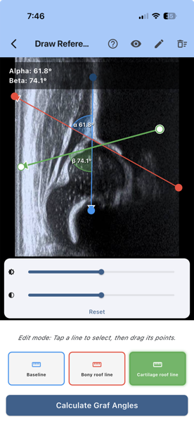
Draw the baseline along the iliac bone, then the bony roof line and the cartilage roof line. Zoom in while you place them.
- 3
Read the alpha and beta angles
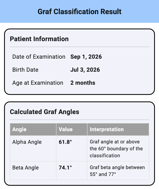
HipAngle Pro works out α and β from your lines and shows them next to the built-in Graf reference guide. Pro adds the Graf type and a PDF report to save or share.
Built around the Graf method
Everything you need to learn, practise and document hip ultrasound measurements.
-
Alpha & beta angles
α and β measured from the three reference lines, with the reference ranges (α ≥ 60°, β ≤ 77°) shown alongside.
-
Graf classification I–IV
Maps the angles you measured onto Graf types I, IIa, IIb, IIc, IId, III and IV, including the age-dependent IIa/IIb distinction — for your own practice and documentation. (Pro)
-
Built-in reference guide
Measurement tips and a Graf reference chart at your fingertips — ideal for training and revision.
-
PDF report & sharing
Generate a clean Graf Hip Classification Report with the image, lines, angles and type. Share by email or any app. (Pro)
-
DICOM receive
Turn your phone into a DICOM receiver on your local network and send images straight from your ultrasound machine or PACS. (Pro)
-
Offline & private
Works fully offline. No account and no backend of ours: images and data stay on your device, with no analytics and no tracking. A report leaves your phone only if you share it yourself.
Who it's for
- Paediatric orthopaedic surgeons
- Radiologists and sonographers
- Paediatricians
- Residents and students learning the Graf method
Available in 15 languages
English, Deutsch, Français, Italiano, Español, Ελληνικά, Polski, Magyar, Hrvatski, Română, Русский, Slovenščina, Srpski, Türkçe, Bosanski.
HipAngle Pro plans
Drawing the lines and reading the angles is free. Pro adds the Graf type classification, PDF documentation and DICOM.
- Graf type classification (I, IIa, IIb, IIc, IId, III, IV)
- PDF Graf Hip Classification Report
- Share reports by email or any app
- DICOM receive from your ultrasound system
Monthly, yearly, or a one-time lifetime purchase — Google Play shows the price for your country. The yearly plan includes a 1-week free trial, which Google Play offers only to users who have never subscribed to this app. Subscriptions auto-renew until cancelled — cancel anytime in Google Play.
Medical disclaimer
HipAngle Pro is intended exclusively as an educational tool to support understanding of the Graf method for developmental dysplasia of the hip (DDH). It is designed for use by trained healthcare professionals or students in a supervised clinical or academic environment.
The visualisations, measurements and classifications shown in the app are provided for training and documentation support only. They are not intended for use in clinical diagnosis or patient treatment.
- This is not a medical device and has not been reviewed or approved by the FDA, EMA or any regulatory authority.
- All calculations are for educational reference only and must not be used to inform or guide real-world clinical decisions.
- Any measurement depends on image quality and on correct anatomical line placement, which may vary across users.
- Final diagnostic and treatment decisions must be made by licensed healthcare professionals using approved clinical tools.